
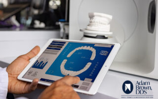
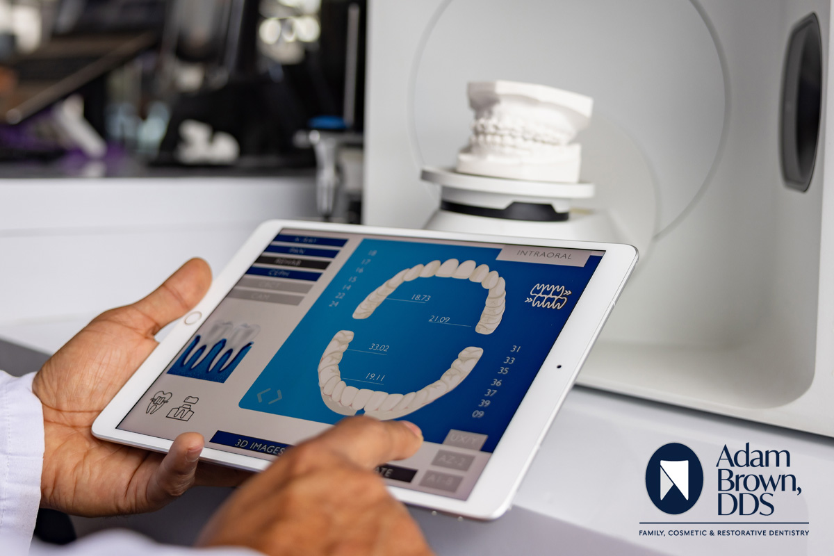
3D Imaging Device
New 3D Planmeca Imaging Device at Adam Brown, DDS
- This 3 dimensional digital device combines three different types of 3D data with one X-ray unit.
- 3D Imaging limits our patients’ exposure to radiation.
- Instead of using standard X-ray film, 3D digital images are produced on the computer screen where we can view them, store them or print them. 3D imaging takes a fraction of the time and uses less radiation.
The X Factor
Wilhelm Conrad Röntgen (1845-1923) was a German mechanical engineer and physicist, who, in 1895 produced and detected electromagnetic radiation in a wavelength range known as X-rays or Röntgen rays, an achievement that earned him the first Nobel Prize in Physics in 1901. (Wikipedia). He demonstrated that the rays could pass through human tissue, yet not bone and teeth. Bone and teeth would appear as “shadows” and he learned to make images from these. Like Pierre Curie, Röntgen refused to patent his discovery, instead wanting society as whole to benefit from his work, he even donated his Nobel Prize money to his university.
X-rays are a form of energy, similar to light and radio waves. X-rays are also called radiation. Unlike light waves, x-rays have enough energy to pass through your body. As the radiation moves through your body, it passes through bones, tissues and organs differently, which allows a radiologist to create pictures of them. The views these images are on photographic film or on monitors similar to a computer display.*
X-ray examinations provide valuable information about your health and help your doctor or dentist make an accurate diagnosis.*
The use of x-rays in dentistry of a living person in the United States took place in 1896. Advances in dentistry and the availability of the equipment grew and x-rays became part of the normal dental routine in the 1950s.
3-D Imaging
3-D imaging has become more popular as machines have become more advanced and more available in the medical community. Popular for ultrasounds, mammograms, and other uses, they are helping doctors and dentists better diagnose health issues, while making it easier on the patient. This is truly technology changing lives.
Dental CBCT (3-D) systems have been sold in the United States since the early 2000s and are increasingly used by radiologists and dental professionals for various clinical applications including dental implant planning, visualization of abnormal teeth, evaluation of the jaws and face, cleft palate assessment, diagnosis of dental caries (cavities), endodontic (root canal) diagnosis, and diagnosis of dental trauma. (www.fda.gov)
Also known as, dental cone beam computed tomography (CBCT), and described by as “a special type of x-ray equipment used when regular dental or facial x-rays are not sufficient. Your doctor may use this technology to produce three dimensional (3-D) images of your teeth, soft tissues, nerve pathways and bone in a single scan.” Adding, “this procedure requires little to no special preparation.” *
More about Cone Bean CT:*
- Cone beam CT is not the same as conventional CT. However, dental cone beam CT can be used to produce images that are similar to those produced by conventional CT imaging.
- With cone beam CT, an x-ray beam in the shape of a cone is moved around the patient to produce a large number of images, also called views. CT scans and cone beam CT both produce high-quality images.
- Dental cone beam CT was developed as a means of producing similar types of images but with a much smaller and less expensive machine that could be placed in an outpatient office.
- Cone beam CT provides detailed images of the bone and is performed to evaluate diseases of the jaw, dentition, bony structures of the face, nasal cavity and sinuses. It does not provide the full diagnostic information available with conventional CT, particularly in evaluation of soft tissue structures such as muscles, lymph nodes, glands and nerves. However, cone beam CT has the advantage of lower radiation exposure compared to conventional CT.
At Adam Brown, DDS, we use Planmeca 3-D imaging device to help diagnose our patients. Not all damage and disease is visible during a routine dental examination. We strive to limit our patients exposure to radiation. That’s why we make the process quick and painless!
The 3-D Planmeca imaging device is the newest X-ray procedures. Instead of using standard X-ray film, 3-D digital images are produced on the computer screen where we can view them, store them or print them. 3-D imaging take a fraction of the time and uses less radiation.
This technology offers up a profound representation of anatomy, thus offering new possibilities for diagnosis and treatment. These advances assist with many forms of dentistry, including: endodontics, periodontics, orthodontics, implantology, dental and maxillofacial surgery.
The benefits for the patient include:*
- Cone beam CT scans provide more information that conventional dental x-ray, allowing for more precise treatment planning.
- CT scanning is painless, noninvasive and accurate.
- A major advantage of CT is its ability to image bone and soft tissue at the same time.
- No radiation remains in a patient’s body after a CT examination.
- X-rays used in CT scans should have no immediate side effects.
- Non-invasive, there is no need to bite down on a mold or piece of plastic.
Benefits for the dentist include:*
- The focused x-ray beam reduces scatter radiation, resulting in better image quality.
- A single scan produces a wide variety of views and angles that can be manipulated to provide a more complete evaluation.
- Surgical planning for impacted teeth.
- Accurate placement of dental implants.
- Determining bone structure and tooth orientation.
- Locating the origin of pain.
- Planning orthodontic issues.
What can you expect?
Like traditional x-rays, you’ll be asked to sit very still. While seated, the x-ray source and detector will sweep around you in unison providing a 360-degree rotation (or less, as needed). This typically takes 20-40 seconds for a full scan, and less if the scan if for a specific area only.
Jewelry, eyeglasses, dentures, and other metal objects may affect the images and should be removed in advance. You might also be asked to remove hearing aids and any removable dental work and piercings.
This is a painless procedure and results are quickly available for treatment planning.
The 3-dimensional digital device at Adam Brown, DDS combines three different types of 3-D data with one X-ray unit! This will save our patients time and additional discomfort associated with traditional X-ray units. As with all procedures, we’ll work with you and determine if your dental insurance covers this technology.
*Radiological Society of North America, Inc.
How Are We Doing?

Schedule Your Next Visit
Blog Posts About Dental Trends

7 Red Flags That Mean It’s Time to Find a New Dentist
Choosing a dentist is an important decision that affects both [...]
2026 Predictions in Dentistry – AI Could Transform Dentistry From Diagnosis to Better Smiles
In a recent 60 Minutes interview, Google DeepMind CEO Demis [...]
 How Artificial Intelligence Can Help You Maintain Dental Health and Save Money at the Dentist Gallery
How Artificial Intelligence Can Help You Maintain Dental Health and Save Money at the Dentist
Adam Brown DDS, Dental Trends, Dentist Office Monroe NC, Oral Health, Preventative Dentistry, Teeth Cleaning, Toothbrush Hygiene
How Artificial Intelligence Can Help You Maintain Dental Health and Save Money at the Dentist
We hear about artificial intelligence (AI) almost every day — [...]